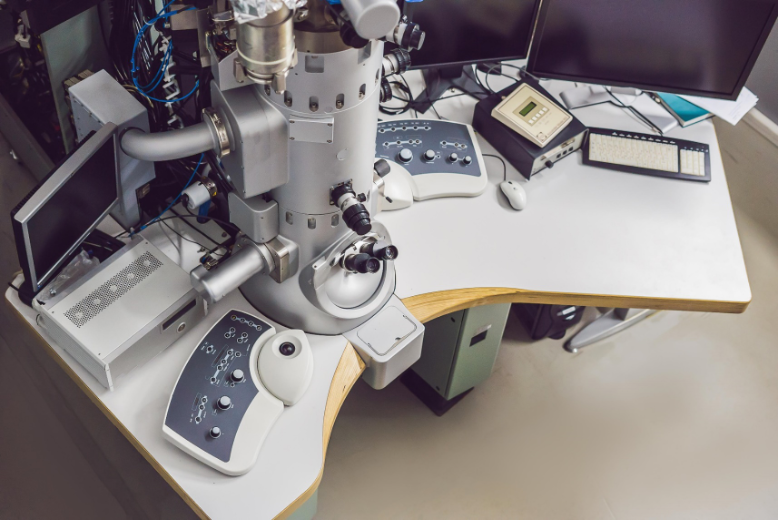
As a high-end scientific instrument, a transmission electron microscope (referred to as a transmission electron microscope) can easily observe the internal structure of a sample, which is at least tens of thousands of times smaller than the smallest structure that can be observed with an optical microscope. It is widely used in fields such as materials science, semiconductor and life sciences. These application fields are characterized by using different types of transmission electron microscopy. In the field of materials science, tungsten filament & lanthanum hexaboride transmission electron microscopy, field emission transmission electron microscopy and spherical aberration transmission electron microscopy are mainly used for characterization. Semiconductors are mainly characterized by Highly automated field emission transmission electron microscopes and spherical aberration transmission electron microscopes are used, while the life science field mainly uses tungsten filament & lanthanum hexaboride transmission electron microscopes and field emission cryo-transmission electron microscopes.
Among them, life science is a basic natural science that is attracting great attention globally today. It is a science that studies the nature, characteristics, occurrence and development rules of life phenomena and life activities, as well as the interrelationship between various biological life activities and the environment. Transmission electron microscopy has been able to expand the microscopic observation of life sciences to the nanoscale level, greatly promoting research and development in the field of life sciences. This article also mainly focuses on the application of transmission electron microscopy in the field of life sciences. The following is an example.
Botany mainly studies the morphology, distribution, genetics, evolution, etc. of plants. The purpose of its research is to better utilize, develop, transform, and protect plant resources. The growth of plants is closely related to human life. In recent years, more and more scientists have used transmission electron microscopy to observe and study the structure of plant tissue cells such as pollen, stems, leaves, roots, and fruits. For example, the changes in internal structure during the morphological development of plants, the differentiation and growth and development processes of tissue cells, exploring the relationship between plant structure and corresponding functions, etc. During the growth of plants, they are often harmed by some pathogens. For example, the leaf tissue of the plant becomes necrotic, causing leaf spots to appear; the conductive tissue of the plant is infected by pathogens, causing the plant to wil and wither; the chlorophyll synthesis metabolism is destroyed, Causes plants to become chlorotic, etc. The use of transmission electron microscopy can conduct microscopic research on botany and pathogens, explore phenomena and patterns in botany, and then help humans improve the disease resistance of plants. It can provide a basis for improving cultivation techniques for economic crops and increasing yields. .
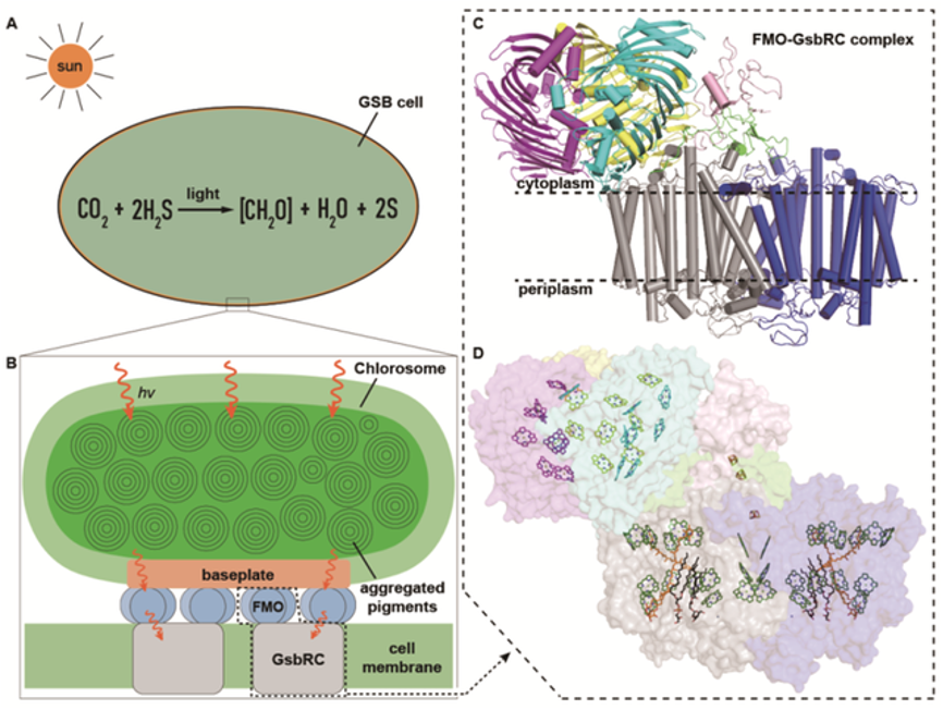
As we all know, photosynthesis is important to both humans and plants. Photosynthesis refers to the process in which green plants use photosynthetic pigments such as chlorophyll to absorb light energy under the irradiation of visible light, convert carbon dioxide and water into energy-rich organic matter, and release oxygen. Photosynthesis has three major functions: first, it can convert energy and convert solar energy into chemical energy; second, it can turn inorganic matter into organic matter and complete material transformation; third, it can regulate the atmosphere and promote the carbon and oxygen balance of the biosphere. In 2020, the Institute of Botany of the Chinese Academy of Sciences and Zhejiang University collaborated to use cryo-transmission electron microscopy to analyze the atomic structure of the photosynthetic reaction center of green sulfur bacteria. This result is of great scientific significance for further exploring the evolution of the photosynthetic reaction center and has been published in top international journals. In the magazine Science. Moreover, in the future, scholars are expected to increase crop yields and alleviate increasingly prominent food and energy problems by artificially simulating the photosynthesis mechanism and bionicly designing photosensitive devices; transforming plant light reaction systems and improving solar energy utilization.

Green sulfur bacteria photosynthetic system and structural model of inner peripheral light-harvesting antenna-reaction center complex (Source: Article mentioned in the text)
Ultrastructural diagnosis is also called electron microscopy diagnosis. Diseases in organisms will lead to morphological and functional changes in cells. With the help of transmission electron microscopy, the morphology analysis and three-dimensional reconstruction analysis of cells in the diseased area can provide a strong basis for disease diagnosis. Avoid misdiagnosis and missed diagnosis, help people diagnose diseases early and treat them correctly. In kidney diseases, due to the unique characteristics of ultrastructural lesions in various kidney diseases, the pathological diagnosis of kidney biopsy is more complicated than that of other diseases. Electron microscopy, immunofluorescence, and light microscopy are indispensable components of kidney biopsy. part. In particular, the role of transmission electron microscopy in the diagnosis of glomerular diseases has received more and more attention. Through transmission electron microscopy, the thickness and structure of the glomerular basement membrane, as well as the shape of the foot processes of its epithelial cells, etc. can be observed. These are important diagnostic basis. At the same time, transmission electron microscopy also plays a pivotal role in ultrastructural diagnostics for studying diseases, distribution and incidence.
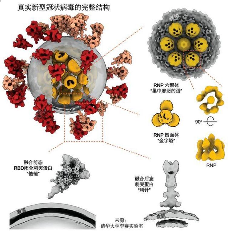
As a pathogen, viruses are usually small in size, and traditional optical microscopes are often difficult to meet the needs for morphological observation. High-resolution transmission electron microscopy has become an important means of current virology research, which can be used to discover viruses and study their characteristics. The structure and composition also provide an intuitive basis for the morphology of the virus. In the hot topic "New Coronavirus Pneumonia" in recent years, transmission electron microscopy has played a role in helping humans observe the morphological identification of the new coronavirus. In the laboratory, the inactivated new coronavirus can be seen clearly under a cryo-transmission electron microscope. The research group of researcher Li Sai from the School of Life Sciences of Tsinghua University and the research group of academician Li Lanjuan of the State Key Laboratory of Infectious Disease Diagnosis and Treatment at the First Affiliated Hospital of Zhejiang University School of Medicine worked closely together to successfully analyze the novel coronavirus (SARS) using cryo-electron microscopy tomography and sub-tomogram average reconstruction technology. -CoV-2) three-dimensional structure of the whole virus. This important research result was published in an internationally authoritative forum on September 15, 2020, Beijing time, under the title "Molecular architecture of the SARS-CoV-2 virus" Published online in the academic journal Cell. This study is the first to analyze the high-resolution molecular structure of the entire novel coronavirus, bringing the world one step closer to understanding the novel coronavirus and helping to further design drugs to block viral invasion. Transmission electron microscopy helps humans understand the microscopic world of viruses and provides support for human scientific research and daily life services.

Real diagram of the internal and external structure of the new coronavirus (source: Li Sai)
With the development of the field of life sciences, cryo-TEM will play an increasingly important role. Cryo-TEM is based on the traditional transmission electron microscope technology, equipped with a low-temperature transmission system and a freezing anti-pollution system. The birth of cryo-TEM has a revolutionary impact on research in the field of life sciences. The Royal Swedish Academy of Sciences awarded the 2017 Nobel Prize in Chemistry to three scholars who invented the cryo-electron microscope: Joachim Frank, a professor at Columbia University, and Richard Henley, a biologist and biophysicist at the University of Cambridge in the United Kingdom. Richard Henderson and Jacques Dubochet, professor emeritus of biophysics at the University of Lausanne in Switzerland. The Nobel Prize Selection Committee said, “Scientific discoveries are often based on the successful visualization of the microscopic world invisible to the naked eye. However, for a long time, existing microscopy technology has been unable to fully display the entire process of the molecular life cycle. Many gaps were left in the biochemical map, and cryo-electron microscopy brought biochemistry into a new era."

From left, Jacques Dubochet, Joachim Frank and Richard Henderson
The main users of cryo-TEM are universities, life science research institutions, hospitals, etc. The demand in China has been increasing in recent years. In terms of production, the current cryo-TEM market is relatively concentrated and is almost completely monopolized by European, American and Japanese manufacturers. Our country has not yet realized the localization of this product, and cryo-TEM will have great room for development in our country in the future.
Global cryo-EM market sales reached US$380 million in 2021, and sales are expected to reach US$590 million in 2028, with a compound annual growth rate (CAGR) of 5.7% (2022-2028). The market value is US$730 million in 2021 and is expected to grow to US$1.41 billion by 2029.

Source: Zhiyin Global Project Database.
In 2009, Tsinghua University began to vigorously develop cryo-EM research and purchased Asia's first cryo-EM. Later, two Titan model cryo-electron microscopes were purchased. Currently, Tsinghua Cryo-EM platform has more than ten pieces of equipment, including four 300kV high-end equipment.
In 2018, the Cryo-EM Center of Southern University of Science and Technology was officially established. The total project investment of the center is 393 million yuan. It is planned to install 6 300kV cryo-EM, 2 200kV cryo-EM, 2 120kV EM, a total of 10 cryo-TEM and 71 others. It has set/sets of related auxiliary instruments and sample preparation equipment, becoming one of the three largest cryo-electron microscopy centers in the world.
In 2021, the multi-modal cross-scale biomedical imaging facility project purchased 5 sets of cryo-EM, and the Shandong University Cryo-EM Center construction project purchased 2 sets of cryo-EM. In 2021, China will purchase a total of 10 sets of cryo-EM, with a total amount of approximately 310 million yuan. In 2022, China's purchase volume of cryo-EM will increase to 17 units, with a total amount exceeding 700 million yuan.
In the past ten years, many universities and scientific research institutions, including the Institute of Biophysics of the Chinese Academy of Sciences, Shanghai Protein Center, Peking University, Fudan University, Shanghai University of Science and Technology, Sichuan University, and Zhejiang University, have introduced high-end cryo-EM. China already has more than 60 cryo-EMs, and Chinese scientists have also achieved world-renowned scientific research results through cryo-EM. For example, scientist Professor Cheng Yifan used cryo-EM technology for the first time to analyze the structure of membrane proteins with near-atomic resolution; Hunan Normal University Professor Liu Hongrong of the University and Professor Cheng Lingpeng of Tsinghua University collaborated to reveal the internal three-dimensional structure of the virus... But from the perspective of cryo-EM manufacturing, China does not yet have the ability to manufacture cryo-EM.

As a high-end scientific research instrument and equipment, transmission electron microscopy strengthens research and development and achieves independent innovation, which meets the needs of the country's long-term development. As the country develops and grows, the requirements for scientific research conditions gradually increase, and the application of transmission electron microscopy will gradually become more widespread, such as using transmission electron microscopy to develop semiconductor chips, carbon nanotubes, graphene, lithium batteries...especially cryo-TEM technology and resolution The improvement in efficiency has overcome the vacuum environment, electron irradiation and other problems of traditional electron microscopy technology, and has greatly promoted human exploration of unknown areas in life sciences.
The 14th Five-Year Plan clearly states that "formulate and implement strategic scientific plans and scientific projects, promote the optimal allocation and resource sharing of scientific research forces in scientific research institutes, universities, and enterprises. Strengthen the research and development and manufacturing of high-end scientific research instruments and equipment." In response to the current international trade war and technical barriers, Chinese companies should strengthen technological innovation in scientific research instruments and equipment, break the technology monopoly of developed countries, and realize the localization of transmission electron microscopes as soon as possible!